Knee pain in your thirties or forties is almost universally attributed to one of two explanations: early arthritis or general wear and tear. Patients are advised to rest, lose weight, take anti-inflammatory medication, and accept that this is what ageing feels like. For some of them, that advice is partially correct. For many others, the actual cause of their knee pain is a completely different condition that responds to a completely different treatment — and they spend years managing the wrong diagnosis. As a leading orthopedic doctor in Kharadi, Dr. Mandar Puranik at orthoclinicpune.com sees this pattern regularly. A proper diagnosis changes everything.
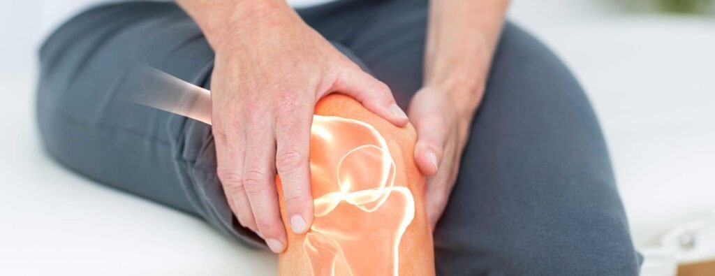
The knee is one of the most structurally complex joints in the body, with multiple distinct components that can each fail independently and produce overlapping symptoms. Pain, swelling, stiffness, and difficulty with stairs are not specific to any one diagnosis — they are generic responses to joint stress that could originate from cartilage, ligament, meniscus, tendon, bursa, or bone. Distinguishing between them requires clinical expertise and the right investigations, not a default assumption. Find Dr. Mandar Puranik on Google to book your evaluation.
Ready to consult? Book An Appointment with Dr. Mandar Puranik today.
The Anatomy of Knee Pain — Why It Is Rarely Simple
The knee joint contains the femur (thigh bone), tibia (shin bone), and patella (kneecap). These surfaces are covered by articular cartilage — a smooth, shock-absorbing layer that allows frictionless movement. Between the femur and tibia sit two C-shaped pads of fibrocartilage called the menisci, which distribute load and provide stability. Four major ligaments — the ACL, PCL, MCL, and LCL — control the joint’s range of motion and prevent abnormal movement. The patella is connected to the quadriceps above and the patellar tendon below, forming the extensor mechanism that allows the knee to straighten.
Any one of these structures can be the primary source of pain. And because they all share a small anatomical space, injury or degeneration in one structure often produces symptoms that mimic injury or degeneration in another. This is why clinical examination by a specialist, combined with appropriate imaging, is essential for an accurate diagnosis.
Conditions That Are Frequently Mistaken for Arthritis
1. Meniscus Tear
A meniscus tear is one of the most commonly misdiagnosed knee conditions in patients over 35. The medial meniscus — the inner pad — is particularly vulnerable to degenerative tears that occur without a specific injury event. The patient simply notices increasing knee pain, stiffness, and sometimes a catching or locking sensation when bending the knee.
Because degenerative meniscus tears occur gradually and are associated with age, they are frequently attributed to arthritis on initial assessment. An X-ray — which shows bone but not soft tissue — will appear normal or show only mild arthritic changes, reinforcing the misdiagnosis. An MRI scan is required to visualise the meniscus clearly. A significant meniscus tear that is causing mechanical symptoms — locking, giving way, pain with specific movements — responds very differently to treatment than arthritis and often benefits from arthroscopic surgery.
2. ACL Insufficiency
The anterior cruciate ligament (ACL) is the primary stabiliser of the knee against forward and rotational forces. A partially torn or chronically insufficient ACL causes persistent instability — a feeling that the knee will give way during cutting movements, descending stairs, or uneven ground. This instability is often described by patients as weakness or unreliability rather than pain, which means it is not always connected to a ligament problem.
In active patients, unaddressed ACL insufficiency leads to progressive damage to the menisci and articular cartilage because the joint is absorbing forces it cannot properly control. Treating the instability early prevents this secondary damage.
3. Patellofemoral Pain Syndrome
Pain at the front of the knee, particularly during stair climbing, sitting for prolonged periods, or squatting, is often labelled as early arthritis when the actual cause is patellofemoral pain syndrome — a condition related to the tracking and mechanics of the kneecap rather than cartilage degeneration. It is significantly more common in women and in patients with certain lower limb alignment patterns. The treatment is primarily physiotherapy targeting hip and quadriceps strength, very different from arthritis management.
4. Pes Anserine Bursitis
The pes anserine bursa sits on the inner side of the knee just below the joint line. When it becomes inflamed — pes anserine bursitis — it causes pain on the inner aspect of the knee that is worse at night, when climbing stairs, and when the knees press together during sleep. This condition is particularly common in overweight patients with osteoarthritis and is frequently attributed entirely to the arthritis when the bursa is actually the dominant pain generator. Targeted treatment of the bursitis produces rapid and significant pain relief.
5. Iliotibial Band Syndrome
Pain on the outer side of the knee that worsens with activity — particularly in runners, cyclists, and people who climb stairs frequently — is often the iliotibial band rather than the joint itself. The IT band is a thick band of connective tissue that runs from the hip to the outer knee. When tight or inflamed, it produces lateral knee pain that is activity-related and reproducible. This is a soft tissue condition, not a joint condition, and responds to stretching, physiotherapy, and activity modification rather than joint-directed treatment.
The most important question in knee pain diagnosis is not how severe the pain is. It is which specific structure is producing the pain. That question can only be answered with a thorough clinical examination and appropriate imaging.
Ready to consult? Book An Appointment with Dr. Mandar Puranik today.
When Is Knee Pain Actually Arthritis?
Genuine knee osteoarthritis is the progressive loss of articular cartilage that covers the joint surfaces. It is a real and common condition, particularly in patients over 50, and it produces a characteristic pattern of symptoms: pain that is worse with activity and better with rest, morning stiffness that improves within 30 minutes of movement, gradual loss of range of motion, and bony enlargement of the joint that is palpable on examination.
An X-ray of a truly arthritic knee shows narrowing of the joint space as cartilage is lost, bony spurs (osteophytes) at the joint margins, and increased density of the bone beneath the cartilage (subchondral sclerosis). These findings, combined with the clinical picture, establish the diagnosis. The severity of arthritis on X-ray does not always correlate with the severity of symptoms — some patients with moderate X-ray changes have severe pain, while others with significant radiological arthritis are relatively comfortable.
The Role of MRI in Knee Diagnosis
An MRI is the investigation of choice when the diagnosis is unclear from clinical examination and X-ray. It provides detailed imaging of all soft tissue structures — cartilage, menisci, ligaments, tendons, and bursae — as well as bone marrow changes that are not visible on X-ray. An MRI does not replace clinical examination; it supplements it. The findings must be interpreted in the context of the patient’s symptoms and examination findings, not in isolation.
At Dr. Mandar Puranik’s orthopedic clinic in Kharadi, every new knee pain patient undergoes a thorough clinical assessment before any imaging is ordered. The clinical examination often narrows the diagnosis significantly and determines which investigations are actually necessary rather than ordering a blanket panel of tests.
Frequently Asked Questions
My X-ray shows mild arthritis but my pain is severe. Which should I trust?
Trust your symptoms. X-ray findings of mild arthritis do not explain severe pain, which means there is likely another pain generator — a meniscus tear, bursitis, or soft tissue problem — that the X-ray is not showing. An MRI and specialist clinical examination will identify it.
Q: Can a meniscus tear heal without surgery?
Some meniscus tears — particularly small, stable tears in the outer zone of the meniscus where blood supply is better — can heal with conservative management including physiotherapy and activity modification. Larger tears, tears that cause mechanical symptoms like locking or giving way, and tears in the inner avascular zone generally do not heal and benefit from arthroscopic treatment.
Q: At what age is knee replacement considered?
Age is not the primary criterion. Knee replacement is considered when arthritis is severe enough to significantly impair quality of life, when conservative treatment has been exhausted, and when the X-ray findings are consistent with the clinical picture. It is performed successfully in patients ranging from their late forties to their eighties depending on the clinical situation.
Years of Knee Pain With No Clear Answer? It Is Time for a Specialist Evaluation.
Dr. Mandar Puranik at Ortho Clinic Pune provides thorough knee assessments that go beyond the standard X-ray and generic diagnosis.
Visit orthoclinicpune.com or find us on Google to book your consultation.
Ready to consult? Book An Appointment with Dr. Mandar Puranik today.Google: Dr. Mandar Puranik on Google | Arthroscopy: Arthroscopy Surgery Kharadi | About: About Dr. Mandar Puranik